Légende
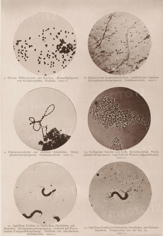
Six microscope images of bacteria and eubacteria, page, photomechanical print, number: 'Tafel II.', inscription: '7. Gross Micrococci from Faeces. Klatschpräparat von Gelatineplatte. Fuchsin. 1000:1. 8. Diplococci in pneumonischem, rostfarbenen Sputum. Deckglassrockenpräparat. Gentianaviolett. 1000:1. 9. Diplococcenkette aus cariöser Zahnhöhle. Deckglassrockenpräparat. Gentianaviolett. 1000:1. 10. Gelbgrüne Sarcine from Luft. Gelatin culture. Deckglasrockenpräparat, ungeefärbt in Wasser eingeschlossen. 1000:1. 11. Spirillum Undula in faulendem Strohinfus, mit Geisseln. Deckglassrockenpräparat. Gebeizt mit Ferrotannat-Campecheholzlösung. Gefärbt mit alkalischem Anilinfuchsin. 1000:1. 12. Spirillum Undula in faulendem Strohinfus, mit Gessel-büscheln. Preparation who bei No. 11. 100:1.', photographer: Carl Günther, Berlin, maker: Friedrich Julius Klinkhardt, Leipzig, c. 1880 - in or before 1890, Saxony, paper, collotype, photoprint, height 242 mm x width 165 mm, Prints in section at the back., unicellular animals, bacteria, bacilli, A vertical sheet showing a grid of circular microscope images arranged in pairs down the page, each circle containing different small organic shapes and textures in sepia tones; the top row shows two circles with scattered dark specks and branching vein-like lines respectively, below them two circles with a looping chain of dots over a darker cluster and a dense honeycomb-like cluster of small rounded units, and the next row of circles features curved wormlike forms against a pale background and a similar single undulating form with radiating thin filamentous appendages; faint printed captions appear beneath each pair of circles.
Crédit
Photo12/Liszt Collection
Notre référence
LZT26B09_289
Licence
Droits gérés
Format disponible
40.9Mo (8.4Mo) / 26.6cm x 38.5cm / 3144 x 4548 (300dpi)