Légende
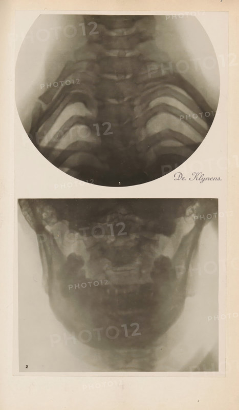
X-ray of an upper and lower jaw, photograph, page, name: 'Dr. Klynens.', c. 1904 - in or before 1909, photographer, paper, gelatin silver print, developing gelatin silver print, height 89 mm x width 103 mm, Photo in section at the back of the book., X-ray examination, jaw (human), A vertical sheet showing two grayscale circular and rectangular radiographic images of a head and upper chest area mounted on a light background, the top image is a round radiograph showing a frontal view of the upper spine and rib outlines with soft gradients of gray and dark curved bone structures, and the lower rectangular radiograph shows a frontal view of the lower skull and jaw with blurred bone contours and symmetric darker areas at the sides, both images bordered by a pale mat and surrounded by an off-white page.
Crédit
Photo12/Liszt Collection
Notre référence
LZT26B07_052
Licence
Droits gérés
Format disponible
40.2Mo (5.0Mo) / 24.2cm x 41.5cm / 2863 x 4902 (300dpi)