Sujet
electron micrograph (TEM) revealing some of the ultrastructure morphology exhibited by a number of different microorganisms
Légende
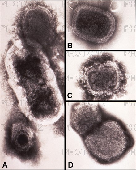
High magnification of 150,000X, negatively-stained transmission electron micrograph (TEM) revealing some of the ultrastructure morphology exhibited by a number of different microorganisms. Panel 'A' represents a composite micrograph, for comparing the size difference between a poxvirus at the top, a bacillus in the middle and a herpesvirus at the bottom. Panels 'B', 'C' and 'D' are TEMs depicting the sequential degeneration of variola virus patricles.
Date
20e siècle
Crédit
Photo12/Ann Ronan Picture Library
Notre référence
ARP18A28_258
Licence
Droits gérés
Format disponible
60.0Mo (2.1Mo) / 34.7cm x 43.3cm / 4100 x 5115 (300dpi)